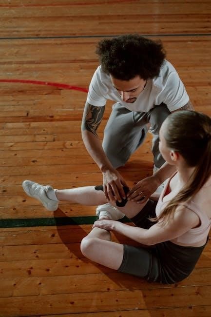
Knee special tests are crucial for diagnosing injuries, evaluating stability, and assessing range of motion; clinical examination guides effective treatment plans.
Accurate assessment relies on understanding test origins and validity, ensuring reliable diagnostic information for orthopedic specialists and informed patient care.
Importance of Accurate Knee Assessment
Precise knee assessment is paramount in orthopedic practice, directly influencing diagnostic accuracy and subsequent treatment strategies. A thorough evaluation, utilizing specialized tests, allows clinicians to pinpoint the specific structures involved in a knee injury – ligaments, menisci, or cartilage – guiding appropriate interventions.
Misdiagnosis or inaccurate assessment can lead to delayed or inappropriate treatment, potentially worsening the patient’s condition and hindering recovery. Understanding the original descriptions and scientific validity of these tests, as highlighted in orthopedic literature, is vital.
Reliable findings from tests like the patellar glide, anterior drawer, and McMurray’s tests contribute to a comprehensive understanding of the knee’s biomechanics and pathology, ultimately optimizing patient outcomes and restoring function.
Overview of Common Knee Injuries
Common knee injuries encompass a wide spectrum of conditions, frequently involving ligament sprains or tears – particularly the ACL, MCL, and PCL. Meniscal tears are also prevalent, often resulting from twisting motions, causing pain, locking, and instability. Patellar instability, including subluxation and dislocation, affects patellofemoral joint function.
Cartilage damage, such as chondral defects, can lead to osteoarthritis, characterized by pain and limited range of motion. Tendonitis and bursitis contribute to localized discomfort. Accurate diagnosis, facilitated by special tests, is crucial.
Effective management requires identifying the specific injury, its severity, and any associated complications, enabling tailored rehabilitation programs and, when necessary, surgical intervention to restore knee stability and function.

Patellar Stability Tests
Patellar stability tests assess the integrity of medial and lateral restraints, evaluating for potential subluxation or dislocation through specific gliding and apprehension maneuvers.
Patellar Glide Test
The Patellar Glide Test assesses patellar tracking and the integrity of the medial and lateral patellar restraints. Performed with the patient supine and the knee flexed to 30 degrees, the examiner gently glides the patella both medially and laterally.
A key indicator involves observing the amount of patellar movement. If the patella glides laterally more than 75% of its width, it suggests laxity in the medial restraints. Conversely, limited gliding – less than 25% – may indicate tightness of the lateral restraints;
This test helps identify potential instability or malalignment contributing to patellofemoral pain syndrome or predisposition to patellar dislocation. Careful observation and comparison to the contralateral knee are essential for accurate interpretation.
Patellar Apprehension Test
The Patellar Apprehension Test evaluates a patient’s subjective sense of stability and potential for patellar dislocation. With the patient supine and the knee extended, the examiner applies a gentle lateral force to the patella, attempting to displace it.
A positive test is indicated by the patient exhibiting apprehension, resistance, or a feeling that their patella is about to dislocate. This sensation, rather than actual dislocation, is the primary endpoint. The examiner should stop applying force immediately if the patient expresses significant discomfort.
This test is particularly valuable in assessing individuals with a history of patellar instability or those suspected of having compromised patellar tracking. It helps gauge the patient’s perceived vulnerability to dislocation.
Patellar Subluxation/Dislocation Test
The Patellar Subluxation/Dislocation Test aims to identify existing or potential patellar instability. The examiner attempts to passively sublux or dislocate the patella, observing the degree of displacement and any associated resistance. This test is performed with the patient relaxed and the knee in a neutral position.
A positive result signifies excessive patellar movement, indicating laxity of the medial patellar restraints. The examiner notes the amount of lateral glide achieved, often correlating it with percentages of the patella’s width. Significant displacement suggests a higher risk of complete dislocation.
Careful observation of patient response and avoidance of forceful manipulation are crucial to prevent further injury or discomfort during this assessment.

Ligamentous Stability Tests
Ligamentous stability tests assess the integrity of the ACL, PCL, MCL, and LCL, crucial for diagnosing knee injuries and guiding rehabilitation.
Anterior Cruciate Ligament (ACL) Tests
ACL testing is paramount in knee injury evaluation, employing several clinical maneuvers to detect laxity or rupture. The Lachman Test, considered highly sensitive, assesses anterior tibial translation with the knee flexed at 20-30 degrees.
The Anterior Drawer Test, performed with the knee flexed to 90 degrees, evaluates anterior tibial displacement, though it may be less sensitive than the Lachman. A positive test indicates ACL insufficiency.
The Pivot Shift Test, a more advanced assessment, aims to reproduce the sensation of giving way experienced during pivoting activities. It assesses rotatory instability combined with anterior laxity, requiring skilled execution and interpretation.
These tests, when combined with patient history and other findings, contribute to an accurate ACL injury diagnosis.
Lachman Test
The Lachman Test is a cornerstone of ACL evaluation, renowned for its high sensitivity in detecting anterior cruciate ligament injuries. The patient lies supine with the knee flexed between 20 and 30 degrees. The examiner stabilizes the femur and applies anterior force to the proximal tibia.
A positive Lachman Test demonstrates increased anterior tibial translation compared to the uninjured side, often accompanied by a soft or mushy end-feel. This indicates ACL laxity or complete rupture.
Careful technique is crucial; excessive force should be avoided. The test assesses the ability of the ACL to resist anterior tibial displacement, providing valuable diagnostic information.
It’s considered more reliable than the anterior drawer test, particularly in acute settings.
Anterior Drawer Test

The Anterior Drawer Test assesses ACL integrity by evaluating anterior tibial translation with the knee flexed to 90 degrees; The examiner stabilizes the foot and pulls the tibia anteriorly, observing for excessive movement relative to the femur.
A positive test reveals increased anterior translation, indicating ACL damage. However, it can be less sensitive than the Lachman test, especially in acute injuries or with hamstring contraction.
The test’s reliability is also affected by patient positioning and muscle guarding. A distinct endpoint should be felt if the ACL is partially intact, while a mushy or absent endpoint suggests complete rupture.
It’s often performed bilaterally for comparison, aiding in identifying subtle differences.
Pivot Shift Test
The Pivot Shift Test is highly sensitive for detecting ACL tears, particularly anterolateral rotatory instability. The patient lies supine with approximately 20-30 degrees of knee flexion and slight internal rotation.
The examiner applies a valgus stress and internal rotation to the tibia while slowly extending the knee. A palpable “clunk” or shift indicates anterior subluxation of the tibia, reduced by the ACL as it tightens during extension.
A positive test signifies ACL insufficiency, as the tibia subluxates anteriorly and then reduces with knee extension. It can be uncomfortable for the patient and requires a skilled examiner.
Grading the test (0-III) helps quantify the severity of instability.
Posterior Cruciate Ligament (PCL) Tests
Evaluating the Posterior Cruciate Ligament (PCL) involves tests assessing posterior tibial translation. The Posterior Drawer Test is performed with the knee flexed to 90 degrees, and the examiner applies a posterior force to the proximal tibia, noting any excessive movement.
The Posterior Sag Sign (Godfrey’s Test) observes the tibia’s resting position with the patient supine and knees flexed. A noticeable posterior sag suggests PCL deficiency.
These tests help identify PCL injuries, often resulting from direct trauma or hyperextension. Accurate assessment is vital for guiding appropriate rehabilitation and potential surgical intervention.
Combined with other clinical findings, these tests contribute to a comprehensive knee evaluation.
Posterior Drawer Test
The Posterior Drawer Test assesses Posterior Cruciate Ligament (PCL) integrity. With the patient supine and the knee flexed to 90 degrees, the foot is stabilized. The examiner then applies a posterior-directed force to the proximal tibia, attempting to translate it posteriorly.
Excessive posterior translation, compared to the unaffected side, indicates PCL laxity or a complete tear. The test relies on feeling for increased movement and observing any palpable step-off.
Proper technique is crucial; hip flexion of 20 degrees helps relax the hamstrings, improving test accuracy. This test is a cornerstone in evaluating PCL injuries resulting from trauma.
Careful interpretation, alongside other clinical findings, is essential for accurate diagnosis.
Posterior Sag Sign (Godfrey’s Test)
Godfrey’s Test, or the Posterior Sag Sign, identifies PCL injuries by observing the tibia’s position relative to the femoral condyles with the patient supine and knees flexed to 90 degrees. A noticeable posterior sag of the tibia, where it appears to fall back compared to the femoral condyles, suggests PCL insufficiency.
This passive test doesn’t require examiner force, relying on gravity to reveal the laxity. It’s particularly useful in acute settings where patient discomfort might limit active testing.
Comparing both legs simultaneously enhances diagnostic accuracy. A positive sign indicates significant PCL damage, often accompanied by instability during weight-bearing activities.
Confirmation requires correlating findings with other clinical evaluations and imaging studies.
Medial Collateral Ligament (MCL) Tests
MCL testing assesses the integrity of the knee’s medial stabilizing ligament. The primary test is the Valgus Stress Test, performed with the patient supine. The examiner applies a valgus force – pushing the knee inward – at varying degrees of flexion (0, 20, and 30 degrees).
Increased gapping on the medial side, compared to the uninjured knee, indicates MCL laxity. Pain during the test also suggests injury, but pain alone isn’t definitive.
Testing at 0 degrees primarily stresses the capsular ligaments, while 20-30 degrees isolates the MCL. Grade I sprains show minimal laxity, Grade II moderate, and Grade III complete rupture.
Careful observation and comparison to the contralateral side are crucial for accurate assessment.
Valgus Stress Test
The Valgus Stress Test evaluates the integrity of the Medial Collateral Ligament (MCL). The patient lies supine, and the examiner applies a valgus force to the knee – pushing inward towards the midline – at 0, 20, and 30 degrees of flexion.
At 0 degrees, the test assesses capsular integrity; 20-30 degrees isolates the MCL. Increased medial joint line opening compared to the uninjured side signifies MCL laxity. Pain indicates potential injury, but isn’t conclusive.
Grading is based on laxity: Grade I (minimal), Grade II (moderate), and Grade III (complete rupture). Proper technique involves stabilizing the hip and ensuring a relaxed quadriceps muscle.
Accurate interpretation requires comparison to the contralateral knee and careful observation for endpoint feel.
Lateral Collateral Ligament (LCL) Tests
Assessing the Lateral Collateral Ligament (LCL) involves evaluating stability against varus stress. The primary test is the Varus Stress Test, performed with the patient supine. The examiner applies an outward force to the knee, attempting to create a varus deformity – bowing outward.
Testing occurs at 0 and 30 degrees of flexion. At 0 degrees, the test assesses combined LCL and capsular integrity; 30 degrees isolates the LCL. Increased lateral joint line opening suggests LCL injury.
Like the MCL test, grading assesses laxity (I, II, III). A firm endpoint is expected; a soft or absent endpoint indicates a more severe injury.
Careful technique and comparison to the uninjured side are vital for accurate interpretation.
Varus Stress Test
The Varus Stress Test specifically evaluates the integrity of the Lateral Collateral Ligament (LCL). The patient lies supine, and the examiner stabilizes the thigh. An externally directed force is applied to the distal aspect of the leg, attempting to create a varus deformity – a bowing outward of the knee.
The test is performed twice: at 0 degrees and 30 degrees of knee flexion. Testing at 0 degrees assesses the combined stability of the LCL and posterolateral corner structures. Flexion to 30 degrees isolates the LCL.
Increased gapping on the lateral side, compared to the uninjured knee, indicates LCL insufficiency. Grading assesses the degree of laxity.

Meniscal Tests
Meniscal tests, like McMurray’s and Apley’s, help identify tears within the knee joint, crucial for diagnosing pain and functional limitations.
McMurray Test
The McMurray Test assesses for meniscal tears by combining knee flexion, tibial rotation, and a palpable click or snap. The patient lies supine, and the examiner fully flexes the knee.
With the heel stabilized, the tibia is rotated internally and externally while applying valgus and varus stress, respectively. A positive test indicates a meniscal tear if a palpable or audible click is felt along the joint line, accompanied by pain.
Sensitivity varies, but it’s a commonly used initial screening tool. False positives can occur, so correlation with other clinical findings is essential for accurate diagnosis and appropriate treatment planning;
Apley’s Compression and Rotation Test
Apley’s Compression and Rotation Test evaluates for meniscal pathology by assessing pain response during compression and rotation of the tibia on a fixed femur. The patient lies prone with the knee flexed to 90 degrees.
The examiner applies axial compression to the tibia while internally and externally rotating it. Pain elicited during rotation, especially with compression, suggests a meniscal tear. The test is then repeated without compression.
Increased pain with compression indicates ligamentous or articular cartilage issues. A positive test is pain reproduction, guiding further diagnostic evaluation and treatment strategies for knee injuries.
Thessaly Test
The Thessaly Test is a highly sensitive clinical examination for detecting meniscal tears, particularly bucket-handle tears. The patient stands on one leg with the knee flexed to 20 degrees, then slowly rotates their body internally and externally.
The examiner observes for any joint line clicking, popping, or a sensation of locking. A positive test is reproduction of joint line pain or a palpable/audible click during rotation, indicating a potential meniscal injury.
This dynamic test stresses the meniscus under weight-bearing conditions, offering valuable diagnostic information alongside other knee assessment techniques for accurate diagnosis.

Range of Motion and Palpation
Assessing knee range of motion and palpating for tenderness or effusion are fundamental steps in a comprehensive knee examination for injury diagnosis.
Knee Range of Motion Assessment
Evaluating knee range of motion (ROM) is a cornerstone of the physical examination, providing vital clues about underlying pathology; Both active and passive ROM should be assessed, noting any limitations or pain experienced by the patient.

Normal knee flexion typically ranges from 0 to 135 degrees, while extension reaches 0 degrees. Observing for deviations from these norms—such as restricted flexion after trauma or hyperextension—is critical.
Documenting the degree of motion achieved during flexion and extension is essential. Furthermore, assess for any crepitus or audible clicks during movement, as these can indicate meniscal tears or articular cartilage damage.

Comparing ROM bilaterally helps identify asymmetries, suggesting unilateral knee pathology. Accurate ROM assessment guides further diagnostic testing and treatment planning.
Palpation for Tenderness and Effusion
Palpation of the knee is a fundamental step in the orthopedic assessment, revealing crucial information about soft tissue and bony structures. Systematically palpate key anatomical landmarks, including the patella, femoral condyles, tibial plateau, collateral ligaments, and joint line.
Assess for localized tenderness, which may indicate ligamentous sprains, meniscal tears, or bony contusions. Carefully evaluate the suprapatellar pouch for evidence of effusion – fluid accumulation within the joint.
Effusion can be detected through a fluid wave or bulge test. Note the temperature of the knee; warmth suggests inflammation. Assess distal pulses and sensation to rule out neurovascular compromise.
Document the location and severity of any tenderness or effusion, as these findings contribute significantly to the diagnostic process and guide subsequent interventions.

Additional Considerations
Comprehensive knee evaluation includes assessing crepitus, joint sounds, and meticulously documenting all findings for accurate diagnosis and effective treatment planning.
Assessing for Crepitus and Sounds
Careful auscultation and palpation during knee examination are vital for identifying crepitus – a grating sensation – or unusual sounds like popping or clicking.
These auditory and tactile findings can indicate various underlying issues, including meniscal tears, articular cartilage damage, or ligamentous instability. The presence, location, and timing of these sounds relative to movement are crucial details.
Documenting the quality of crepitus (coarse, fine) and the specific movements that elicit sounds helps differentiate potential causes. While not always indicative of pathology, abnormal sounds warrant further investigation alongside other clinical findings to formulate an accurate diagnosis and appropriate treatment strategy.
Documentation of Findings
Thorough and precise documentation of all knee examination findings is paramount for effective patient care and communication among healthcare professionals.
Detailed notes should include observations from the range of motion assessment, palpation, ligamentous stability tests, and any special tests performed, noting both positive and negative results. Specific measurements, such as degrees of motion or millimeters of joint line opening, should be recorded accurately.
Clearly describe any crepitus, sounds, or tenderness elicited during the examination. A well-documented examination provides a baseline for tracking progress, guiding treatment decisions, and supporting medicolegal requirements, ensuring continuity of care.